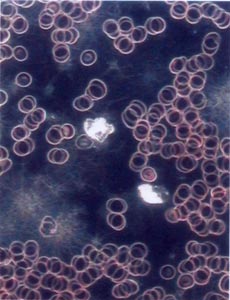
Darkfield Microscopy

Darkfield Microscopy Blood Analysis is an analysis of your body’s internal environment. A single drop of blood from your finger is able to provide a composite of over 25 aspects. Darkfield microscopy now allows us to observe multiple vitamin and mineral deficiencies, toxicity, and tendencies toward allergic reaction, excess fat circulation, liver weakness and conditions that favour arteriosclerosis.
Darkfield Microscopy Blood Analysis is carried out by placing a drop of blood from the patient’s fingertip on a microscope slide under a glass cover slip to keep it from drying out.The slide is then viewed at high magnification with a darkfield microscope that forwards the image to a television screen. This allows both the doctor and patient to view the blood cells.
Blood is magnified by 30,000 times, giving us the ability to assess how the body is compensating to its environment and to monitor your progression while on treatment. Darkfeild Microscopy Blood analysis is not a diagnostic procedure for specific diseases. It is best used to help determine the optimal diet and most effective supplementation (enzymes, herbs, antioxidants, etc.).
Darkfield Microscopy can show:
- The quality of red blood cells
- Free radical damage to the blood cell and the need for antioxidants
- Acid/alkaline balance in the body
- The tendency to develop atherosclerotic plaque and hyper-clotting disorders
- Evidence of bacteria, parasites, Candida /yeast /fungi (indirect measurement)
- Undigested proteins and fats
- Evidence of hormonal imbalances
- Folic acid, B12 and iron deficiencies
- Uric acid crystals and risk for gout
- Poor circulation, oxygenation levels
Article: Darkfield Microscopy – “You canT grow a flower in cement”
